
Artificial Vision History
The initial observations on electrical stimulation of the visual cortex began in Germany at the end of World War I, during the course of neurosurgical operations under local anesthesia. In 1918, it was found that motionless dots of light, later called “phosphenes,” could be seen in the patients’ contralateral visual fields through stimulating the surface of the visual cortex,.
The long-time dream of curing blindness started in the early sixties in England with Dr. Giles Brindley’s research and culminated in 1968 when he implanted in a blind volunteer an electrode array of 80 stimulating electrodes against the visual cortex of the right hemisphere of her brain. He demonstrated that it was possible to transfer external visual information into the patient’s mind, such that she could identify simple patterns and letters from combinations of multiple phosphenes. The results of his work were published in a scientific paper [1] in 1969, which became a landmark in visual research. Brindley’s investigation led to an international conference at the University of Chicago in 1969, aimed at trying to find future directions necessary to explore a possible visual prosthesis. Two paths resulted from the conference: the studies at the NINDS Neuroprosthesis Program to determine the safe limits of chronic cortical stimulation in animals; and human stimulation studies by William Dobelle at the Division of Neuroprosthesis of the Department of Artificial Organs at the University of Utah headed by Dr. Willem Kolff.
Inspired by Brindley’s work, Dr. William Dobelle devoted himself to the research of artificial vision with the aim of designing a visual prosthesis that could eventually restore vision to the blind. In the late sixties and early seventies, William Dobelle and his team, traveling to a number of medical centers in North America, performed acute stimulation experiments on sighted patients who had to undergo brain surgery due to unrelated medical problems, and volunteered along with their surgeons to provide observations arising from such stimulation carried out intraoperatively under local anesthesia.
Subsequent to these studies, which lasted no more than one hour, Dr. John Girvin at the University of Western Ontario in London, Ontario, Canada operated on three blind volunteers recruited from the state of Utah’s Agency for the Blind and performed acute stimulation studies, lasting for several days, after which the electrode arrays were removed. Those studies largely confirmed the findings of Giles Brindley.
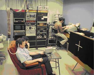
In the late seventies, several volunteers were implanted with chronic implants and the results were very promising. Over a ten-year period, one volunteer provided important observations, being able, for example, to read “phosphene Braille” much faster than “tactile Braille.” He could also identify the orientation of white strips on a black board by manipulating a video camera mounted on a joystick (left)
Since Brindley’s research, the focus was to assist the blind in reading. However, in the eighties the emphasis shifted to independent ambulation, as the development of the cassette tape and the availability of most reading material, and more particularly books on tapes relegated the primary aim of reading to a lesser importance. The need to design a portable device was impeded by the size of the electronics, the amount of current drawn by the batteries and their size. Only in the late nineties, with the advances in electronics miniaturization and computer technology, did it become possible to design a truly portable artificial vision system.
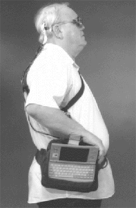
Experiments with the last implanted volunteer yielded more promising results. He was the first patient to use a portable system (right). It consisted of a miniature video camera mounted in the frame of his glasses, connected to a sub-notebook computer which processed the images, and a stimulator which generated the pulses to the visual cortex. A cable connected the external hardware to a percutaneous connector mounted on his skull, and internally connected to the implanted electrode array. He was able to be placed in a room, recognize and retrieve a black ski cap from a far wall, turn around, recognize a mannequin, walk over and place the cap on its head. This led to his recognition in the Guinness Book of Records in 2001.
Between April 2002 and January 2004 sixteen blind patients were implanted in Lisbon, Portugal with an artificial visual prosthesis consisting of two (bioccipital) surface electrode arrays. The results were quite encouraging. Patients could actually “see” objects, walk in the streets, find trees, lamp posts, fire hydrants, walk into the entrance of a building, orient themselves, find doors and other points of reference, grab objects off a table, etc. Even though it had very low resolution – the average number of phosphenes varied between 60 and 100 – most patients were excited with their new “vision”. One patient was able to drive a car around perceived objects in a private parking lot. Although there are researches going on in several centers around the world to try to restore vision to blind people due to retinal diseases, cortical stimulation is the only way to provide vision to blind people with damaged optic nerves. The only limitation is that it does not apply to people born blind or blinded as children. Unfortunately, Dr. Dobelle passed away in 2004. His family would like to keep his legacy to benefit the blind. They have reached an agreement with Stony Brook University on Long Island, New York, to transfer the related intellectual properties for continuous development within the university setting.
[1] Brindley, G.S. and Lewin, W.S. (1968) The sensations produced by electrical stimulation of the visual cortex. J. Physiol., 196, 479-493.
Copyright 2006, All rights reserved.